

Related Topics

Research Explainer & Analysis — This article explains findings from a peer-reviewed study published in Science and provides additional scientific context, interpretation, and discussion of the study’s significance and limitations. The analysis is intended for educational purposes and distinguishes established findings from possible future applications.
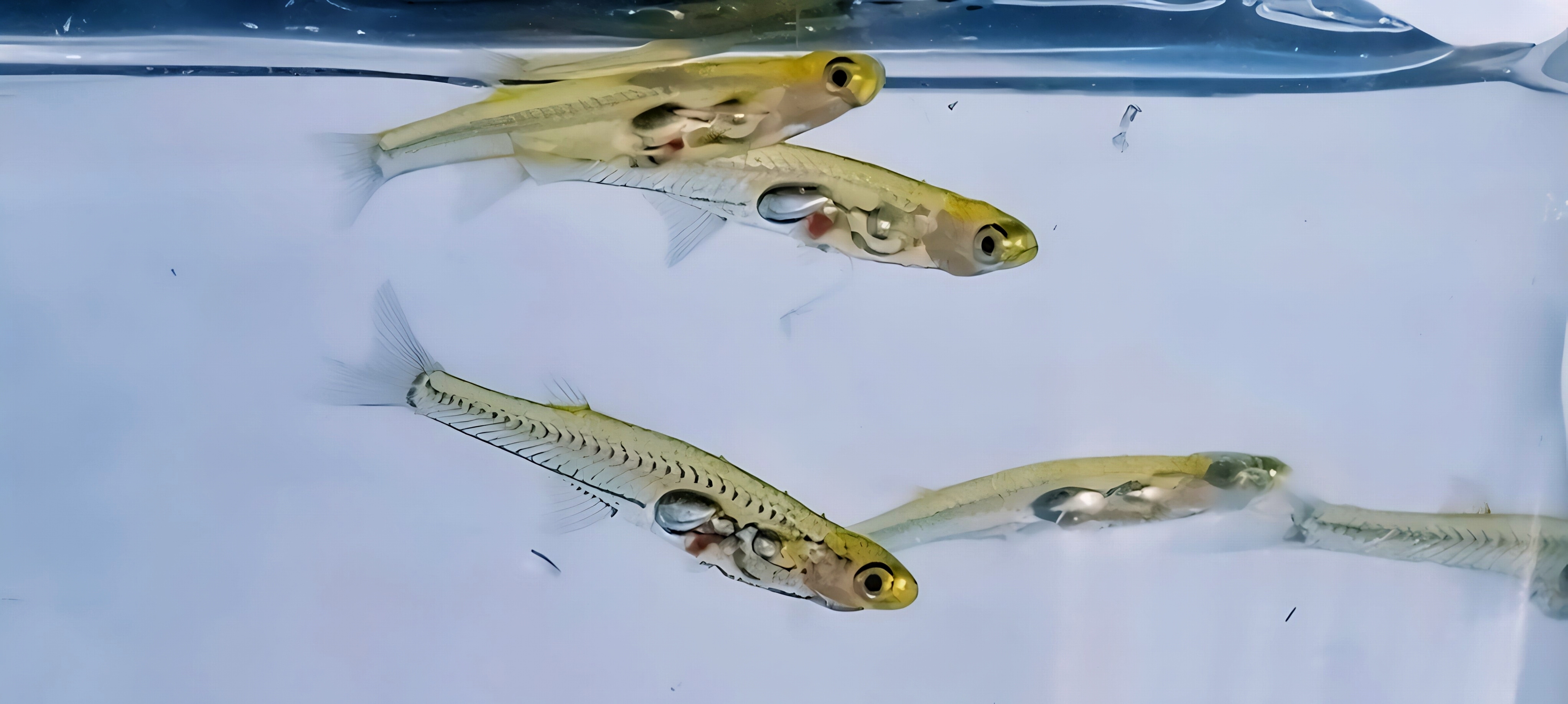
Scientists have created a detailed cellular map of an entire adult vertebrate, offering a new way to investigate how the body’s cells function, maintain their identities, and respond to injuries. In a study published in Science, researchers from the Whitehead Institute for Biomedical Research and collaborating institutions examined more than 600,000 cells from a tiny, nearly transparent fish called Danionella cerebrum. This fish measures approximately one centimeter in length, making it small enough for scientists to study cells throughout its body. Using single-cell RNA sequencing, the researchers identified different cell populations by examining which genes were active inside them. They also used spatial methods to determine where many of these cells were located. The resulting atlas provides a comprehensive reference of the fish’s cellular organization, including its brain, spinal cord, internal organs, and fins. Unlike studies that examine only one organ, this research offers a broader picture of how specialized cells are distributed throughout an adult animal (Atabay et al., 2026).
Why is mapping individual cells important? Although nearly all cells in an animal contain essentially the same genetic instructions, different cells use different combinations of genes to perform specialized functions. For example, nerve cells transmit information, muscle cells produce movement, and immune cells help protect the body. Single-cell RNA sequencing allows researchers to investigate these differences at a molecular level by measuring RNA, which provides information about gene activity. However, an important distinction is that this method generally measures a snapshot of gene expression rather than directly revealing everything a cell does. In the new study, researchers found that neurons located in the same brain region often shared similar gene-expression patterns, even when they used different neurotransmitters to communicate. This suggests that a neuron’s location and developmental history may be important aspects of its identity, alongside its communication function. The researchers also discovered that certain connective tissue cells retained gene-expression patterns associated with the development of specific body regions. These findings raise an interesting biological question: could adult cells retain positional information that helps tissues preserve their structure or rebuild it after injury? The study identifies molecular patterns consistent with this possibility, but further experiments are needed to establish their precise functions (Atabay et al., 2026).
One of the most interesting applications of this cellular atlas involves brain regeneration, an ability that differs considerably among vertebrate species. In humans, significant damage to brain tissue often results in lasting neurological problems because many lost neurons are not readily replaced. However, some fish can generate new nerve cells following certain types of brain injury. The researchers investigated this process in Danionella cerebrum by studying cellular responses following injury to the telencephalon, a region of the forebrain. They observed a sequence of biological changes involving wound responses, immune activity, neural stem cells, and the formation and maturation of new neurons (Atabay et al., 2026). These findings are important because tissue regeneration involves more than simply producing replacement cells. New neurons must develop appropriate identities, occupy suitable locations, and potentially establish functional connections with existing neural networks. The atlas provides a way to identify which cell populations participate in regeneration and which genes may be involved. Nevertheless, the production of new neurons does not automatically demonstrate that every damaged neural connection or lost behavior has been restored. Additional studies examining neural connectivity and functional recovery would help determine how completely the regenerated tissue restores normal brain activity.
What does this discovery mean for humans? The greatest value of this research may lie in the scientific questions it makes possible rather than in any immediate medical application. By comparing the cellular responses of Danionella cerebrum with those of mammals, researchers may be able to identify biological pathways associated with successful tissue regeneration and investigate why similar responses are more limited in humans. For example, future studies could examine whether particular stem-cell populations, immune responses, or developmental gene programs contribute to the formation of replacement neurons. However, identifying similar genes in fish and humans would not necessarily mean that those genes perform identical functions or could safely produce the same regenerative effects. Differences in brain structure, cellular organization, and injury responses must also be considered. Another limitation is that a cellular atlas is primarily a descriptive resource: it identifies cell populations and their molecular characteristics but does not, by itself, prove that specific genes cause regeneration. Experiments that alter gene activity and measure the resulting biological effects will be needed to establish cause-and-effect relationships. The researchers have made their cellular data available through an interactive resource called DigiFish, allowing other scientists to investigate these questions. Ultimately, the study demonstrates how examining an entire small vertebrate at the cellular level can deepen our understanding of development, tissue maintenance, and regeneration, while providing a foundation for future discoveries in neuroscience and regenerative biology (Atabay et al., 2026; White, 2026).
References
Atabay, K. D., Aoude, P., Park, C., Kadobianskyi, M., Paugois, O. H., Judkewitz, B., & Reddien, P. W. (2026). Whole-body single-cell atlas of an adult vertebrate, Danionella cerebrum. Science, 394, eaeg1125. https://doi.org/10.1126/science.aeg1125
White, M. (2026). Researchers create first whole-body single-cell atlas of an adult vertebrate. Whitehead Institute for Biomedical Research. https://wi.mit.edu/news/researchers-create-first-whole-body-single-cell-atlas-adult-vertebrate
AI Disclosure Statement:
The Chromoscience uses AI-assisted tools to support the preparation and editing of science articles. Content is reviewed by the editor for clarity and consistency with the cited research before publication. Readers are encouraged to consult the original scientific publications for complete findings, methods, and limitations.

Leave a Reply