

Related Topics

Scientists now have a clear picture of the secret structure that helps shape a simple multicellular organism. This shows how cells work together to create complex living things.
Scientists from Britain and Germany published a study in the Proceedings of the National Academy of Sciences that showed the organization of the extracellular matrix in Volvox carteri. V. carteri is a species of green algae that is frequently utilized to examine how multicellular organisms transformed from single-celled relatives.
The extracellular matrix (ECM) is a scaffold-like structure that encompasses cells and helps them develop and communicate. Additionally, it shapes the cells and controls how they appear. It is found in animals, plants, fungi, and algae, and it was significant in the change from single-celled to complex life.
Scientists think that the ECM forms through self-assembly, which is still not fully known even in the simplest living things. This is because the ECM appears outside of the cells that make it.
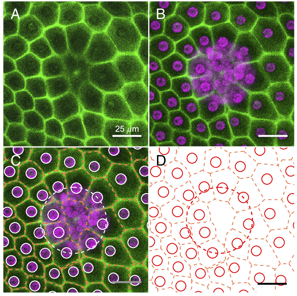
To find out more, scientists at the University of Bielefeld genetically modified a type of Volvox so that a key ECM protein called pherophorin II became fluorescent. This made it possible to see the framework of the matrix extremely clearly using a microscope.
The researchers saw a complex network of rounded components that looked like foam and covered around each of Volvox’s about 2,000 somatic cells, which are not reproductive cells.
The team used machine learning to measure the shape of these sections with the help of scientists from the University of Cambridge. The results showed a growth pattern that was unpredictable. This pattern is similar to how foams expand when they get soaked.
According to statistics, these forms followed a pattern that can be found in living cells as well as grains and emulsions. The results show that even though different cells make ECM proteins at different rates, the organism as a whole stays in a normal, spherical shape.
The fact that single cells can behave in a chaotic way and the whole organism can have precise geometry makes us wonder how complex life manages to make reliable shapes from parts that don’t work well together.
The findings provide insight into a basic developmental biology question: how can cells generate exterior structures reliably? It also shows what amazing things scientists, physicists, and mathematicians can do when they work together to figure out how life works.
By following a single structure protein, the researchers learned how the extracellular matrix organizes itself. The way it looks tells us something useful about how the organism changes as it grows.
Sources:
Benjamin von der Heyde et al. (2025). Spatiotemporal distribution of the glycoprotein pherophorin II reveals stochastic geometry of the growing ECM of Volvox carteri, Proceedings of the National Academy of Sciences. DOI: 10.1073/pnas.2425759122
Glowing algae reveal the geometry of life. https://phys.org/news/2025-08-algae-reveal-geometry-life.html
AI Disclosure Statement:
The Chromoscience uses AI-assisted tools to support the preparation and editing of science articles. Content is reviewed by the editor for clarity and consistency with the cited research before publication. Readers are encouraged to consult the original scientific publications for complete findings, methods, and limitations.



Leave a Reply